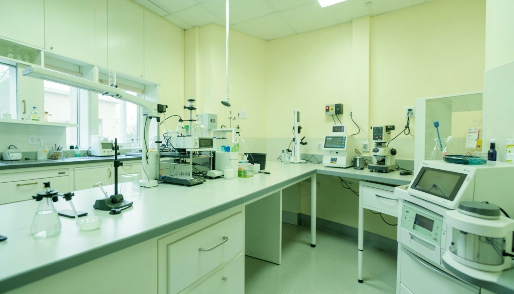
Building Competence in TB Fluorescent Microscopy
Training lab staff on proper use of TB fluorescent microscopy requires more than demonstrating how to operate a microscope. Reliable results depend on a complete workflow: receiving and identifying specimens, preparing smears, applying stains correctly, examining slides systematically, recording findings, maintaining equipment, and escalating problems. Each task affects the accuracy and safety of tuberculosis diagnosis.
A quality-focused program should be adapted to the laboratory’s workload, staffing, equipment, and available supplies. Staff may work with LED fluorescence microscopes, different auramine-based staining procedures, paper registers, or electronic reporting systems. The training approach must therefore define essential practices while allowing controlled adaptation to local conditions.
Competence is best developed through demonstration, supervised practice, direct observation, and documented assessment. A staff member should be able to explain why each step matters, perform it consistently, recognize an unacceptable result, and take appropriate corrective action. The laboratory’s quality management system provides the structure for making that competence routine rather than dependent on individual experience.
Define The Competency Standard
Before training begins, the laboratory should describe what a competent microscopist is expected to do. The standard may include specimen identification, slide labeling, smear preparation, staining, microscope setup, systematic scanning, grading of acid-fast bacilli, result recording, cleaning, and safe waste handling. It should also cover the ability to recognize artifacts, contamination, poor staining, and instrument problems.
Competency statements should be observable and measurable. “Understands fluorescent microscopy” is too broad to assess effectively. A stronger statement might require the employee to prepare a smear of suitable thickness, stain it within the approved procedure, examine the required area without skipping fields, and report results using the laboratory’s authorized terminology.
Training should distinguish initial qualification from ongoing competency. New staff need structured instruction and close supervision, while experienced staff benefit from periodic reassessment, review of discrepant results, and updates when procedures or equipment change. A written authorization list can show which activities each employee may perform independently and which still require supervision.
Prepare A Safe And Consistent Workflow
Good microscopy begins before the slide reaches the microscope. Staff should be trained to verify patient identifiers, specimen type, collection date, request details, and acceptance criteria. Any mismatch or unsuitable specimen should be managed according to a documented procedure rather than resolved informally at the bench.
Biosafety instruction must be integrated into every practical session. TB specimens can generate infectious aerosols during manipulation, so staff need clear guidance on personal protective equipment, work inside the appropriate containment device, spill response, hand hygiene, sharps management, and decontamination. Fluorescent staining does not remove the biological risk associated with an untreated specimen.
The physical arrangement of the work area also influences quality. Clean and potentially contaminated materials should move in a controlled direction, reagents should be clearly identified, and stained slides should be separated from unprocessed specimens. Trainers can use a workflow map to show where labeling, staining, reading, reporting, and disposal occur, then observe whether staff follow it during routine work.
Teach Staining And Slide Examination
Practical instruction should explain the purpose of each staining step rather than reduce the procedure to memorized timings. Staff need to understand how smear thickness, reagent condition, decolorization, drying, and contamination affect fluorescence and interpretation. Reagents should be checked for labeling, preparation date, storage conditions, expiry, and visible deterioration before use.
Microscope training should cover the instrument’s light source, filters, objectives, stage movement, focusing technique, and safe handling. Staff should learn how to set up the field consistently and examine slides in a defined pattern. A systematic scanning method reduces the chance of missing bacilli and makes reading speed easier to monitor.
Reference slides and image sets are valuable teaching tools, but they should supplement, not replace, supervised examination of actual prepared slides. Learners should compare negative material, scanty positives, stronger positives, debris, staining artifacts, and poorly prepared smears. The trainer should discuss why an image is reportable or questionable and how uncertainty is resolved through a second reading or referral.
Verify Performance With Evidence
A written quiz alone cannot establish microscopy competence. Assessment should combine direct observation, practical slide reading, review of recorded results, and discussion of safety and quality control. The assessor should use the same criteria for every employee and retain the evidence in an individual training or competency file.
The following framework can help organize an assessment session:
| Competency Area | Evidence To Review | Example Acceptance Measure |
|---|---|---|
| Specimen and slide handling | Observed workflow and labels | Identifiers match throughout processing |
| Smear preparation | Direct observation of prepared slides | Smear is even, suitable, and correctly labeled |
| Staining | Reagent checks and procedure observation | Steps, timing, and quality checks follow the SOP |
| Microscope operation | Demonstration and instrument checklist | Microscope is set up, focused, and cleaned correctly |
| Slide reading | Blind panel or coded slides | Results meet the laboratory’s defined agreement target |
| Reporting | Completed worksheets or registers | Findings are recorded accurately and promptly |
| Safety and waste | Observation and scenario response | Required controls are used without prompting |
Acceptance limits should be defined before testing. They may include agreement with reference results, maximum reading errors, completion of all critical safety steps, and accurate documentation. If a staff member does not meet the standard, the response should be additional coaching and reassessment, not an informal pass based on familiarity or staffing pressure.
Assessment results should identify the specific gap. A person who reads slides accurately but forgets equipment checks needs a different intervention from someone who struggles to distinguish weak fluorescence from artifacts. Targeted retraining is more efficient and gives supervisors a clearer basis for follow-up.
Record Training And Supervision
Training records should show the employee’s name, dates, topics, trainer, assessment method, results, identified gaps, corrective actions, and authorization status. Records should be legible, protected from loss, and easy to retrieve during internal reviews or external assessments. The GLI Quality Tool’s user instructions can help laboratories navigate its resources and apply them within a broader quality management approach.
A paper-based system can work well when it has defined file locations, consistent naming, controlled forms, and a simple index. Laboratories that rely on paper records can use this guidance on organizing TB lab records to strengthen document retrieval and retention practices. Training evidence should be linked to the employee’s current role and reviewed when responsibilities change.
Supervisors should observe staff after the formal training period. Short, routine checks are often more useful than infrequent intensive inspections. An observation might focus on slide labeling one week, microscope cleaning the next, and result transcription later. Feedback should be immediate, specific, and recorded when it identifies a recurring risk.
Use Quality Data For Improvement
Routine quality indicators can show whether training is producing dependable performance. Useful measures include rejected specimens, mislabeled slides, staining failures, unreadable smears, discordant readings, delayed reports, equipment downtime, and repeated documentation errors. These data should be reviewed for patterns rather than used only to assign blame.
When a problem appears, supervisors should examine the entire process. A weak result may reflect inadequate training, but it may also arise from expired reagents, an unstable power supply, poor ventilation, microscope maintenance problems, excessive workload, or unclear procedures. Corrective action is more effective when it addresses the actual cause.
The GLI Quality Tool organizes improvement work across quality systems essentials and provides a Phase 4 roadmap for laboratories working toward sustained quality. Its approach can support regular review of competency records, quality indicators, corrective actions, and management responsibilities. Fluorescent microscopy training should become part of this continuing cycle rather than a one-time event.
Practical Priorities For Laboratory Managers
A manageable training program can begin with a small number of high-value actions and expand as evidence accumulates. Managers should align the training calendar with service demand, ensure that staff have access to approved procedures, and protect time for supervised practice. The following priorities help establish a dependable foundation:
- Create a role-specific competency checklist covering safety, specimen handling, staining, microscopy, reporting, and equipment care.
- Use coded reference slides and direct observation to assess both technical accuracy and workflow discipline.
- Keep reagent, equipment, quality control, and training records together in a controlled retrieval system.
- Review errors and near misses during regular laboratory meetings, focusing on causes and corrective action.
- Reassess staff after procedure changes, extended absence, new equipment installation, or repeated quality problems.
Managers should also make sure trainers are themselves competent and consistent. Different supervisors should not give conflicting instructions about scan patterns, grading terminology, or reporting rules. A current standard operating procedure, shared assessment materials, and periodic trainer review can reduce variation across shifts.
A strong program gives staff confidence to stop and seek help when a result is uncertain. That behavior protects patients and supports trustworthy surveillance data. When laboratory leaders connect practical microscopy skills with documented quality practices, each slide becomes part of a controlled diagnostic process rather than an isolated technical task.
Begin by mapping the current workflow, selecting the critical competencies, and observing one complete microscopy session. Use the findings to create a focused training and assessment schedule, then record progress and review the results through the laboratory’s quality meetings. Consistent reinforcement will help turn fluorescent microscopy into a safe, reproducible, and sustainable TB diagnostic service.