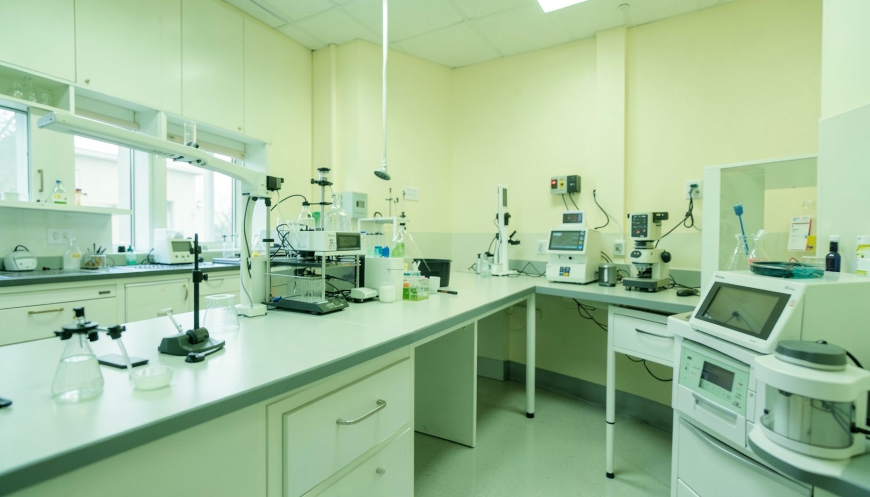
Preventing Cross-Contamination in TB Molecular Testing Workflows
Molecular testing has transformed tuberculosis diagnosis by detecting Mycobacterium tuberculosis complex and identifying important drug-resistance markers quickly. Yet the same sensitivity that makes nucleic acid amplification tests valuable can also expose a laboratory to false-positive results when amplified material, patient specimens, or contaminated equipment move into the wrong area.
Cross-contamination prevention is therefore a process-wide responsibility. It begins before a specimen reaches the molecular platform and continues through extraction, reagent preparation, amplification, result review, waste disposal, and environmental monitoring. A reliable workflow makes clean practices easy to follow and errors difficult to introduce.
Laboratories working in different resource settings may use automated cartridges, manual extraction, line-probe assays, or open-platform PCR. The technology varies, but core principles remain consistent: separate activities, control movement, use appropriate personal protective equipment, document deviations, and investigate unexpected results systematically.
Map the workflow before changing it
Begin by drawing the physical and operational path of a specimen. Mark where samples are received, opened, aliquoted, extracted, amplified, and discarded. Include staff routes, supply delivery, equipment access, waste collection, and the movement of paperwork or electronic records. This exercise often reveals hidden overlaps, such as a staff member carrying clean reagents through a specimen processing area.
A unidirectional arrangement is preferred. Materials should move from clean activities toward progressively more contaminated activities, rather than returning to an earlier stage. In a compact laboratory, complete physical separation may not be possible. In that case, laboratories can use time separation, thorough decontamination, dedicated trays, closed containers, and carefully controlled staff movement.
The workflow map should also identify contamination hazards at each step. Opening high-burden specimens, transferring extracted nucleic acid, handling positive controls, and opening amplified reaction products can all release material that affects later testing. Risk assessment should consider the assay design, room layout, ventilation, equipment, staffing levels, and daily workload.
Keep clean and dirty activities apart
Molecular testing areas should be divided according to function. Reagent preparation belongs in the cleanest area, followed by specimen preparation and nucleic acid extraction. Amplification and post-amplification analysis should be physically separated from pre-amplification work wherever possible. Dedicated equipment, coats, gloves, pipettes, racks, and consumables reduce the chance that contamination will travel between zones.
Pipettes are particularly important. A pipette used for extracted DNA or amplified product should never return to reagent preparation. Filtered aerosol-resistant tips should be used, and tips must be changed between every specimen and control. Operators should avoid splashing, forceful pipetting, and touching tube rims or caps with gloves that have contacted samples.
Staff behavior can undermine even a well-designed layout. Personnel should move from clean areas to dirty areas, not in the reverse direction during the same work session. Gloves must be changed when moving between activities, and hands should be disinfected after glove removal. Doors, phones, keyboards, pens, and shared benches require attention because they can become overlooked transfer points.
Control specimens, reagents, and controls
Specimen reception should include clear identification, leak checks, appropriate packaging, and prompt placement in a designated holding area. Samples should remain closed until they reach the correct processing station. Open tubes should be kept upright, and racks should be stable and easy to disinfect. Any spill, damaged container, or labeling discrepancy requires documented action before testing continues.
Positive controls deserve special handling because they contain target material capable of contaminating future reactions. Prepare and use them in a controlled area, preferably near the end of the setup sequence. Negative controls and no-template controls should be included according to the assay procedure and reviewed before patient results are released. A pattern of unexpected positivity in controls may indicate reagent, environmental, or carryover contamination rather than true patient infection.
Reagent aliquoting can reduce repeated opening of stock containers and limit the effect of a single contamination event. Expiry dates, lot numbers, storage temperatures, and freeze-thaw exposure should be recorded. Calibration and maintenance also support contamination control; a useful calibration schedule guide can help laboratories organize this work when equipment support is limited or distributed across rural sites.
| Workflow point | Main contamination risk | Practical control | Evidence to retain |
|---|---|---|---|
| Specimen receipt | Leaking or mislabeled containers | Inspect, segregate, and document before opening | Receipt and rejection records |
| Sample preparation | Aerosols, spills, and tube-to-tube transfer | Use filtered tips, stable racks, and careful pipetting | Work sheet and incident log |
| Extraction | Carryover between specimens | Change tips, disinfect surfaces, and follow run order | Run file and control results |
| Reagent setup | Introducing target DNA into clean reagents | Use a dedicated clean zone and aliquots | Lot and preparation records |
| Amplification and analysis | Opening amplified products near clean work | Keep post-amplification work isolated | Instrument report and review |
| Waste handling | Spread from used tubes and consumables | Close containers promptly and decontaminate safely | Waste and cleaning records |
Make cleaning and waste control routine
A written cleaning schedule should specify what is cleaned, by whom, with which disinfectant, at what frequency, and how completion is recorded. Work surfaces should be cleaned before and after testing and immediately after spills. The disinfectant must be compatible with the surface and used at the correct concentration and contact time. A quick wipe that evaporates too soon may provide little protection.
Cleaning should proceed from cleaner areas toward dirtier areas, using dedicated materials for each zone. Reusable racks and tools need a defined decontamination process. UV light, where used, should never be treated as a substitute for physical cleaning or chemical disinfection, because its effectiveness depends on exposure, distance, shadowing, lamp performance, and surface condition.
Waste containers should be close enough to prevent carrying open or contaminated materials across the room. Sharps must enter puncture-resistant containers, while tubes, tips, and other infectious waste require appropriate leak-resistant disposal. Procedures should address spills, broken tubes, power interruptions, and instrument faults so that staff can respond consistently rather than improvising during a stressful event.
Detect contamination before it affects reporting
Quality control should look for trends, not just isolated failures. Review negative control results, invalid rates, unexpected positivity, repeat testing, and changes associated with a reagent lot, operator, instrument, or work area. A sudden rise in positive results among low-signal specimens may warrant an investigation even when individual runs appear acceptable.
Environmental monitoring can support an investigation when contamination is suspected. Swabs from benches, pipette stands, instrument surfaces, handles, and other high-touch locations may help identify a source, although the sampling plan must be validated and interpreted carefully. Testing should never be delayed indefinitely by unfocused environmental sampling; the laboratory should define actions, responsibilities, and decision points in advance.
When a contamination event occurs, stop and contain the problem. Preserve run records, identify potentially affected specimens, quarantine relevant reagents, clean the area, and notify the technical supervisor. Determine whether results require repeat testing, correction, or communication to clinicians. The role of external quality assessment also matters because proficiency testing can reveal weaknesses in analytical performance that internal controls do not expose.
Strengthen the system through documentation
A dependable prevention program is built into the laboratory quality management system rather than left to individual memory. Standard operating procedures should describe workflow zoning, specimen handling, pipetting, control placement, cleaning, spill response, waste disposal, equipment maintenance, and result review. Versions must be controlled, accessible at the point of use, and updated when methods or layouts change.
Training should combine explanation with direct observation. New staff need demonstrated competency before independent testing, while experienced staff benefit from periodic reassessment. Supervisors can use checklists to observe glove changes, pipette technique, movement between rooms, control handling, and documentation practices. Corrective coaching should focus on the process and its risk, not simply on assigning blame.
Laboratories can organize these activities within the broader quality framework described in the twelve Quality Systems Essentials. Personnel, facilities and safety, equipment, purchasing, documents, assessment, and continual improvement are connected. A contamination problem may appear to be a pipetting error but actually reflect inadequate training, poor supply control, crowded workspace, or missing supervisory review.
Priorities for a practical prevention program
Resource limitations do not eliminate the need for contamination control, but they do require thoughtful prioritization. Laboratories should first address high-impact risks that can be managed with clear procedures and consistent behavior. Improvements can then be tracked through measurable indicators such as control failures, contamination investigations, rejected specimens, and repeat-test rates.
Useful actions include:
- Separate clean reagent preparation, specimen processing, and post-amplification activities as far as the facility allows.
- Assign dedicated pipettes, racks, protective clothing, and cleaning materials to each workflow zone.
- Use filtered tips, frequent glove changes, stable tube racks, and careful low-aerosol pipetting.
- Review controls and contamination trends before authorizing patient results.
- Record cleaning, maintenance, training, incidents, corrective actions, and follow-up verification.
A strong program is visible in ordinary details: the direction staff walk, where waste is placed, how a positive control is handled, and whether an unexpected result triggers a documented review. These routines protect patients from incorrect diagnoses and help laboratories maintain confidence in their TB molecular results.
Use the GLI Quality Tool to assess the current workflow, identify gaps across the quality system, and turn those findings into practical actions suited to the laboratory’s setting. Start with one mapped process, assign responsible staff, document the change, and review the evidence until contamination prevention becomes a reliable part of everyday testing.