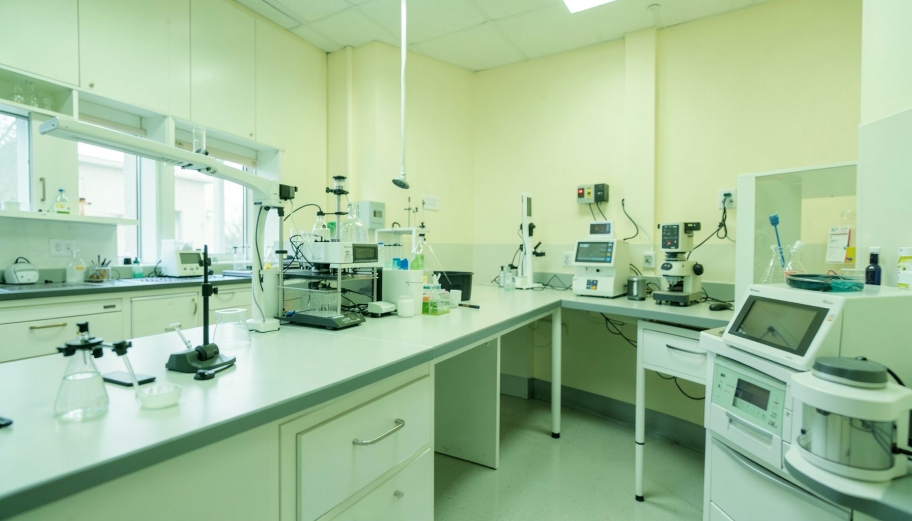
Troubleshooting contamination in TB culture workflows
Contamination in tuberculosis culture can obscure true results, delay treatment decisions, waste scarce specimens, and reduce confidence in laboratory data. It may appear as bacterial or fungal growth on solid media, turbidity or unusual instrument signals in liquid culture, or a rising rate of invalid and contaminated results over time.
A single contaminated tube does not always indicate a laboratory-wide failure. The cause may be introduced before the specimen reaches the laboratory, during processing, through defective reagents or media, or from weaknesses in equipment, workflow, or environmental controls. Effective troubleshooting therefore begins with a defined investigation rather than an immediate change to the decontamination procedure.
A quality management system helps teams connect contamination events with records, staff practices, supplies, maintenance, and trends. The goal is to identify the point at which contamination entered the process, apply a proportionate corrective action, and confirm that the change has worked.
Start with the pattern, not the assumption
Review contamination by specimen type, collection site, referring facility, date, staff member, work shift, test method, media lot, and processing batch. Calculate the contamination rate using a consistent denominator, such as the number of specimens inoculated or cultures set up. Separate contamination in solid culture from contamination in liquid culture because the patterns and detection methods may differ.
A sudden increase across several specimen types may indicate a common problem, such as a contaminated reagent, water source, biosafety cabinet issue, or lapse in cleaning. A cluster linked to one collection site points more strongly toward specimen collection, transport, container integrity, or delays before processing. Sporadic events may reflect individual handling errors, while persistent low-level contamination can signal a process that has gradually weakened.
Keep the original records available during the review. Accession logs, worksheets, instrument printouts, media quality-control results, temperature records, maintenance logs, and nonconformity reports can reveal links that are missed when staff rely on memory. The phase-specific checklists can help organize this review across equipment, personnel, documents, assessment, and process control.
Check the specimen before checking the bench
Pre-analytical problems are a frequent source of culture contamination. Examine whether specimens arrived in sterile, leak-proof containers with secure lids and clear identifiers. Assess transport time and temperature against the laboratory’s validated requirements. Delayed delivery, excessive heat, leakage, or repeated opening of containers can give environmental organisms an opportunity to multiply before processing.
The collection method also matters. Sputum contaminated with saliva, material collected in unsuitable containers, or specimens handled in crowded areas may carry a larger microbial burden. Review instructions provided to patients and collection staff, and check whether collection sites have adequate supplies, privacy, hand hygiene facilities, and a reliable route for prompt transport.
When contamination is concentrated in specimens from one location, communicate the findings without assigning blame. A short review with the collection and transport teams can identify practical causes, such as reusable transport boxes that are not cleaned correctly, specimens held over weekends, or labels and requisitions that encourage unnecessary handling.
Examine reagents, media, and decontamination steps
Culture media and reagents should be checked for correct preparation, storage, expiry, labeling, and lot traceability. Inspect containers for unexpected turbidity, precipitation, color change, damaged seals, or evidence of repeated temperature excursions. Review sterility and growth-promotion quality-control results before releasing a new lot for routine use.
The decontamination process must balance removal of unwanted organisms with preservation of mycobacteria. Excessive exposure to the decontaminating agent, incorrect concentration, inaccurate timing, or poor neutralization can reduce recovery of Mycobacterium tuberculosis complex. Insufficient treatment can leave too many non-target organisms alive. Staff should follow the approved, validated standard operating procedure rather than making informal adjustments.
Compare actual practice with the documented method. Verify pipettes, timers, centrifuges, and measuring devices where relevant, and observe whether staff use the correct order of additions, mixing technique, and processing volumes. Small variations can become important when several steps are performed manually or when workload is high.
| Pattern observed | Likely areas to investigate | Useful checks |
|---|---|---|
| Contamination rises after a new reagent or media lot | Preparation, storage, sterility, supplier, lot handling | Review lot records, QC results, expiry dates, and retained samples |
| Several specimens in one batch are contaminated | Batch setup, workspace, shared reagents, equipment | Compare worksheets, staff assignments, cabinet cleaning, and reagent use |
| Contamination is linked to one collection site | Collection quality, containers, transport, holding time | Review collection instructions, transport logs, and site feedback |
| Liquid culture contamination increases while solid culture remains stable | Liquid-system handling, bottle septa, instrument workflow | Check inoculation technique, bottle integrity, instrument alerts, and controls |
| Persistent contamination across all work areas | Environment, water, cleaning, ventilation, workflow separation | Assess surfaces, cabinet certification, cleaning records, and room practices |
| Contamination falls after a staff change or retraining | Technique, adherence, supervision, workload | Observe procedures and compare competency records |
Review the workspace and equipment
Work areas should support a one-directional workflow from cleaner activities toward more hazardous or contaminated materials. Unnecessary movement, open containers, overcrowded benches, shared pens, and poorly separated clean and dirty supplies increase opportunities for cross-contamination. Observe the process at busy times, since shortcuts often appear when specimens arrive in large batches.
Check that the biological safety cabinet is appropriate for the work, used correctly, and certified at the required intervals. Cabinet alarms, damaged surfaces, blocked grilles, poor sash position, or excessive equipment inside the cabinet can compromise containment and work quality. Cleaning and disinfection should follow a written schedule that identifies the product, concentration, contact time, responsible person, and response to spills.
Centrifuges, racks, vortex mixers, pipettes, incubators, and liquid culture instruments also deserve attention. Look for cracked tubes, leaking buckets, aerosols, residue, incorrect balance, unstable temperatures, or overdue preventive maintenance. A recurring contamination pattern after centrifugation, for example, warrants an observed review of tube loading, rotor condition, sealing, and cleanup rather than a general reminder to “be careful.”
Use controls and records to locate the failure
Quality-control results can help distinguish contaminated material from a contaminated process. Uninoculated media controls, reagent controls, and other locally defined controls should be interpreted according to the laboratory’s procedures. A positive control may show that a medium can support growth, but it does not prove that the entire workflow is free from contamination. Negative controls and environmental monitoring, where validated and appropriate, can provide additional evidence.
When contamination is detected, record enough detail to reconstruct the event. Include the specimen identifier, date and time, staff involved, batch or lot numbers, equipment used, culture system, observed appearance, and immediate action. Photographs may be useful where permitted by policy. Avoid deleting or rewriting original records; corrections should remain traceable.
Trend data should be reviewed at a frequency suited to workload and risk. A monthly summary can show whether contamination is stable, increasing, or concentrated in a particular subgroup. Laboratories should define alert or action thresholds in their quality documents and escalate findings according to the potential effect on patient results.
Turn findings into corrective action
An investigation should end with a specific root cause or a clearly documented range of plausible causes, not simply “staff error.” Ask why the error was possible. Was the SOP unclear? Was the workspace poorly arranged? Were supplies unavailable? Did staffing, fatigue, equipment failure, or training gaps contribute? A systems-based review produces stronger preventive action than retraining alone.
Immediate actions may include quarantining a suspect media lot, checking affected batches, notifying supervisors, reviewing potentially impacted results, or arranging repeat testing when clinically appropriate. Follow the laboratory’s reporting and referral procedures, especially when contamination may have delayed detection of drug-resistant TB or affected a critical patient result.
Corrective actions should have an owner, deadline, required resources, and a method of verification. The team might revise specimen transport instructions, repair a centrifuge, change the movement of materials, strengthen lot acceptance testing, or introduce direct observation of a high-risk step. Building a continuous improvement culture helps make these actions part of routine laboratory learning rather than a response limited to one incident.
Build contamination control into assessment and review
Contamination prevention works best when it is embedded in daily management. Supervisors can use brief process observations, review control results during team meetings, and discuss near misses before they become reportable failures. Staff should be able to raise concerns about supplies, workload, workflow, or equipment without fear that every problem will be treated as individual misconduct.
Internal audits should test whether procedures are followed in practice, whether records are complete, and whether previous corrective actions remain effective. An external assessment can provide an independent view of biosafety, quality control, document management, and continual improvement. Preparing systematically for a WHO SLMTA assessment can also help a TB laboratory connect contamination control with broader quality system requirements.
Practical priorities for a laboratory beginning this work include:
- Establish a contamination log with consistent definitions and review it routinely.
- Separate pre-analytical, analytical, and post-analytical causes during every investigation.
- Verify media, reagent, equipment, and environmental controls before changing validated procedures.
- Use direct observation and competency assessment for critical manual steps.
- Document corrective actions and confirm their effectiveness with follow-up trend data.
When contamination is treated as a measurable quality signal, the laboratory can move from repeated troubleshooting to prevention. Each well-documented event strengthens specimen handling, culture reliability, staff confidence, and the safety of the testing environment. Use the findings from the next contamination review to assign clear actions, monitor the trend, and reinforce a culture in which reliable TB results are everyone’s responsibility.