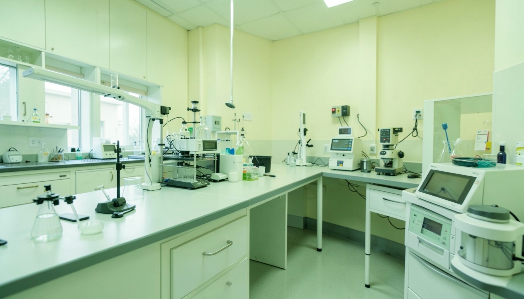
Designing a faster TB smear microscopy workflow
Turnaround time in tuberculosis microscopy is shaped by every handoff between receiving a specimen and releasing a result. Delays may arise from accessioning, slide preparation, staining, drying, reading, result verification, or reporting rather than from the microscope itself. A faster workflow therefore depends on removing avoidable waiting while protecting smear quality and patient safety.
The goal is a predictable sequence that allows staff to begin useful work as soon as a specimen arrives. This includes clear acceptance criteria, visible work queues, appropriate batching, defined responsibilities, and a reliable process for urgent or rejected samples. Speed should be measured alongside error rates, contamination, slide quality, and the proportion of results reported within the laboratory’s target time.
A quality management system gives the workflow structure. The GLI Quality Tool can help TB laboratories connect operational changes with documentation, personnel competency, equipment management, assessment, and continual improvement, including in settings where staffing and supplies are limited.
Map the current specimen journey
Begin by documenting the actual path of a sputum specimen, from arrival at the facility to communication of the result. Record who receives it, how it is labeled, where it waits, when it enters the register, how slides are prepared, and how results move from the reader to the clinician. Observing the process during busy and quiet periods often reveals queues that staff may not notice during routine work.
Measure elapsed time at each step rather than recording only the final turnaround time. Useful timestamps include specimen receipt, registration, smear preparation, staining, microscopy start, result verification, and report release. A simple process map can distinguish active work from idle time caused by missing forms, unavailable stains, shared equipment, or the absence of an authorized reviewer.
Separate specimens by urgency and processing needs. A routine diagnostic sample, a follow-up sample, and a specimen from a hospitalized patient may require different reporting priorities. The laboratory should define these categories in writing, while ensuring that urgent handling never bypasses identification checks, biosafety precautions, or result verification.
Standardize reception and accessioning
A rapid microscopy service begins with a controlled reception point. Staff should check the patient identifiers, specimen type, volume, collection date, request form, and container condition immediately on arrival. Samples that meet acceptance criteria should be registered without being placed in an unmonitored holding area.
Rejection rules should be practical and consistent. When a sample is unsuitable, staff need a documented reason, a clear route for clinician notification, and a procedure for recollection. Correcting an identification problem early is faster and safer than preparing a slide that later has to be discarded or investigated.
Use a specimen log or laboratory information system that shows status at a glance. Entries such as “received,” “in preparation,” “staining,” “awaiting reading,” and “reported” make bottlenecks visible. If electronic systems are unavailable, a wall board or paper batch sheet can provide the same operational control, provided it is protected from unauthorized access and updated at defined times.
Arrange work around flow, not habit
The physical layout should follow the order of operations. Receiving, smear preparation, staining, microscopy, verification, and reporting areas should be clearly separated, with materials positioned near the step where they are used. This reduces unnecessary movement and helps limit cross-contamination between clean and potentially infectious activities.
Prepare supplies before the work period begins. Slides, applicators, labels, staining reagents, timers, absorbent material, personal protective equipment, and waste containers should be checked against a short opening checklist. A missing marker or expired reagent can stop an entire batch, so preparation time is often a better investment than trying to work faster during microscopy.
Batching can improve efficiency when specimens arrive steadily, but excessive batching creates hidden delay. Laboratories should define a maximum waiting period before preparation begins. A small batch may be appropriate during low-volume periods, while a larger service can use scheduled preparation cycles with an exception pathway for urgent specimens.
| Workflow point | Common source of delay | Practical control | Useful indicator |
|---|---|---|---|
| Reception | Incomplete forms or unclear identifiers | Immediate acceptance check and rejection log | Time from arrival to registration |
| Smear preparation | Shared bench or missing supplies | Dedicated workstation and opening checklist | Time from registration to slide preparation |
| Staining | Long unattended batches | Validated batch size and timed steps | Percentage of slides stained within target |
| Microscopy | Slides waiting for an available reader | Assigned reading blocks and queue board | Time from staining to reading |
| Verification | Reviewer unavailable | Planned review periods and escalation route | Time from reading to authorization |
| Reporting | Manual transcription or delayed communication | Standard report format and release log | Time from verification to report |
Protect slide quality while increasing speed
Reducing turnaround time must never mean shortening validated staining steps, skipping drying, or reading poorly prepared smears. A technically weak slide can produce a false-negative or an unreliable grading result, creating a much greater delay through repeat testing and clinical investigation.
Standard operating procedures should define the smear size and thickness, drying conditions, staining method, reagent preparation, quality control, and microscope maintenance. Each critical step should have an observable acceptance criterion. For example, staff can check that a smear is evenly distributed, fully dry, correctly labeled, and free of visible defects before staining.
Microscopy reading should be organized to reduce interruptions and fatigue. Readers need adequate light, a clean and correctly adjusted microscope, appropriate oil where required, and a defined field-reading method. Scheduled reading blocks are generally more consistent than asking staff to examine slides between unrelated duties.
A visual reference can support consistent interpretation among readers, especially when grading scanty, low-positive, or high-bacillary-load smears. The visual grading guide describes how a laboratory can create a local aid for standardizing smear grading and training personnel.
Define roles, checks, and escalation
Every stage should have an assigned owner and a backup person. Reception staff may verify identifiers and register samples, a designated technician may prepare and stain slides, and an authorized reader may examine and release results. Clear role boundaries prevent samples from waiting simply because staff assume someone else is responsible.
Use a second-person check at points where errors have serious consequences, particularly patient identification, result transcription, positive findings, and report authorization. The check should be quick and documented. It should verify the original slide and worksheet rather than relying only on a copied result.
Escalation rules keep exceptions from disappearing in the queue. A specimen that has exceeded its target time, a broken microscope, a failed stain control, or a discrepant reading should trigger a defined response. The procedure may include notifying the supervisor, moving work to a backup area, repeating quality control, or referring the specimen according to local policy.
Competency assessment should reflect the real workflow. New and reassigned staff need direct observation, review of prepared slides, and documented authorization before independent reading. Refresher training can focus on the specific error patterns or delays identified through monitoring rather than repeating every procedure equally.
Monitor turnaround time and improve continuously
Set a measurable service target, such as the percentage of eligible specimens reported on the day of receipt or within a defined number of working hours. The target should reflect local transport schedules, staffing, biosafety requirements, and clinical needs. A target that cannot be achieved consistently will not guide improvement effectively.
Review turnaround time by stage and by shift. Median time can describe typical performance, while the longest times reveal exceptional failures. Also monitor rejected specimens, repeat smears, unreadable slides, quality control failures, transcription errors, and corrective actions. These measures show whether a faster process is actually more reliable.
The laboratory can test one change at a time, such as moving registration closer to reception, introducing a morning supply check, or reserving a daily reading period. Compare results before and after the change, discuss unintended effects with staff, and update the relevant procedure if the change is retained. The Phase 4 guidance is relevant when a laboratory is using monitoring, corrective action, and continual improvement to sustain better performance.
Actions that make the workflow dependable
A practical implementation sequence can help a laboratory move from observation to controlled change:
- Draw the current specimen pathway and record waiting time at every handoff.
- Establish written acceptance, rejection, prioritization, and escalation criteria.
- Create a visible queue that shows each specimen’s current status.
- Use an opening checklist for supplies, equipment, stains, safety materials, and workspace readiness.
- Review turnaround indicators with staff at a fixed interval and document corrective actions.
The best design will vary with specimen volume, staffing, transport arrangements, and available technology. A small laboratory may gain the most from disciplined reception and scheduled reading, while a larger service may need separate preparation and reporting queues. In both settings, the essential principle is the same: make each step visible, assign responsibility, and prevent avoidable waiting.
A well-designed TB smear microscopy workflow is a controlled system rather than a race. When laboratories combine sensible batching, reliable slide preparation, competent reading, rapid verification, and regular performance review, they can shorten reporting time without weakening diagnostic quality. Start by timing the current process for one representative week, then use the findings to implement one documented improvement at a time.